Why your heel hurts with the first steps
Heel pain often comes from strain and inflammation in the plantar fascia. This strong band under your foot supports its arch and absorbs shock as you walk. A common sign is sharp pain with your first steps in the morning or after sitting for a long time. It may ease as you move, then return after prolonged standing [1, 2, 6]. Repeated strain can cause tiny injuries where the band joins the heel bone. The area can then feel irritated and stiff after rest or sleep [4, 6].
At La Rose Clinics in New Maadi and the Fifth Settlement, Dr Shaimaa Sherif, a specialist in rheumatology, joint disorders, physical medicine and rehabilitation, assesses heel and foot pain with an examination and ultrasound. This helps distinguish repeated strain from inflammatory tendon problems. You can Book your appointment to discuss your symptoms and care plan. This guide covers causes of plantar fasciitis, the role of heel spurs, stretches and other first treatments, and signs that need prompt rheumatology care.
What can make heel pain more likely?
Plantar fasciitis rarely develops in isolation; rather, it emerges from cumulative mechanical loading that outpaces the ability to heal of the tissue. The American Academy of Orthopaedic Surgeons and the National Health Service identify several major physical risk factors [1, 2, 6]:
1. Excess Body Weight: Elevated body mass imposes direct, unyielding mechanical compressive and tensile forces on the plantar arch during every step; research indicates high body mass index is one of the strongest predictors of recalcitrant plantar fasciopathy [6, 10]. If you are seeking a safe strategy to relieve lower-limb joint stress, explore our guide discussing weight loss and knee osteoarthritis, detailing how body mass reduction decreases compressive force across lower extremity structures. 2. Occupational Prolonged Standing: Professions requiring sustained weight-bearing on hard floors, such as teachers, retail staff, surgeons, and hospitality workers operating on concrete or tiled floors [1, 2]. 3. Structural Foot Morphologies: A rigid high arches clearly reduces physiologic shock absorption, concentrating impact directly upon the heel tuberosity, whereas flat feet causes excessive strain and elongation along the fascial band [2, 6]. 4. Calf Muscle and Triceps Surae Tightness: Contracture of the calf muscles and Achilles tendon limits ankle upward movement, compelling the midfoot and plantar fascia to absorb abnormal compensatory tensile loads during walking [2, 10]. 5. Inadequate Footwear: Wearing unsupportive flat shoes, flimsy sandals, or walking barefoot on hard domestic tile floors, alongside worn-out athletic footwear lacking shock-absorbing midsoles [1, 4]. 6. Sudden Increases in Physical Activity: Abrupt escalation in walking volume, running mileage, or sudden high-impact interval workouts without structured progression [2, 6].
Does a heel spur cause the pain?
One of the most pervasive medical misconceptions surrounding heel pain involves the heel spur, frequently referred to in popular parlance as a heel nail. When a patient experiencing severe heel pain undergoes a routine X-ray, the X-ray often reveals a small pointed bony projection protruding from the inferior calcaneus. Patients frequently assume this spur acts like a physical spike digging into soft tissue with every step [2, 7, 9].
However, Imaging and tissue studies show that this view is incorrect: 1. The Bone Spur Does Not Cause the Pain: A study published in Foot and Ankle Surgery by Johal and Milner evaluating large patient cohorts found that heel spurs are findings present in 15% to 20% of people without symptoms who have never experienced heel pain [9]. Conversely, a substantial proportion of people with severe plantar fasciitis demonstrate completely normal heel morphology with no spur on an X-ray [7, 9]. 2. How Do Spurs Actually Form? The spur is a secondary natural response instead of a main cause. Chronic mechanical tensile traction across the bone surface stimulates gradual calcium deposition and bone spur formation over months and years, functioning as a structural adaptation instead of a sharp foreign object [2, 7]. 3. Surgical Spur Removal is Obsolete: Contemporary guidelines strongly discourage surgical excision of the heel spur. Historical procedures aimed at resecting or shaving the spur frequently destabilized the arch of your foot while failing to address the true pain generator, which is wear and inflammation within the plantar fascia [7, 9].
Consequently, successful clinical management focuses on relieving fascial tension, restoring flexibility, and eliminating abnormal mechanical strain, instead of targeting an asymptomatic bony spur observed on imaging [2, 6].
When heel pain may be linked to rheumatism
While the majority of heel pain presentations represent mechanical overuse, a vital clinical subset reflects peripheral enthesitis secondary to systemic axial or peripheral spondyloarthritis (SpA). In spondyloarthritis, systemic immune dysregulation targets the enthesis—the specialized anatomical junction where tendons, ligaments, and fascial bands insert into bone [3, 8, 11].
The where it joins the heel bone of the plantar fascia and the posterior insertion of the Achilles tendon represent the two primary anatomical sites for lower-limb enthesitis [3, 12]. Recommendations from the European Alliance of Associations for Rheumatology (EULAR) highlight that high-resolution musculoskeletal ultrasound with Power Doppler provides indispensable clarity, detecting increased blood flow where the tendon joins bone and early bone changes that reliably distinguish inflammatory tendon attachment pain from isolated mechanical wear [11, 12].
A assessment by a rheumatologist is essential if chronic heel pain coexists with any of the after other symptoms: 1. Prolonged Morning Stiffness: Heel pain or peripheral joint stiffness persisting longer than 30 to 60 minutes upon waking, which paradoxically improves with continuous moderate physical movement and worsens after inactivity or prolonged rest [3, 8]. 2. Inflammatory Spinal Symptoms: Deep, unyielding lower back or buttock ache awakening the patient during the second half of the night, relieved by warm showers and gentle ambulation; this is the clinical hallmark of ankylosing spondylitis affecting axial sacroiliac joints and spine [8, 11]. 3. Cutaneous and Nail Involvement: Well-demarcated erythematous psoriatic plaques on the elbows, scalp, or umbilicus, alongside nail pitting, onycholysis, or dactylitis (sausage digits), suggesting psoriatic arthritis targeting entheseal structures [3, 8]. 4. Extra-Articular Inflammatory Pathology: Episodes of acute anterior uveitis characterized by painful photophobia and scleral erythema, or chronic digestive symptoms pointing toward inflammatory bowel disease [3, 8, 11].
What else can cause heel pain?
When evaluating a patient presenting with persistent heel pain, clinicians must thoroughly exclude alternative lower-extremity pathologies that closely mimic plantar fasciitis but need distinct treatment pathways [4, 5, 6]:
1. Calcaneal Stress Fracture: Resulting from sudden escalations in impact loading or underlying osteopenia. The critical diagnostic difference is that stress fracture pain steadily accelerates throughout weight-bearing activity instead of easing after initial morning steps, and elicits sharp local pain during medial-lateral manual heel compression (the heel squeeze test) [5, 6]. 2. Peripheral Nerve Entrapment and Tarsal Tunnel Syndrome: Compression of the posterior tibial nerve or its first branch (Baxter's inferior heel nerve) produces paresthesias, electric shooting sensations, burning numbness radiating across the plantar aspect, and pain that frequently worsens during nocturnal recumbency [5, 6]. 3. Achilles Tendinopathy and Retroheel Bursitis: Symptoms localize primarily to the posterior heel margin, roughly 2 to 4 centimeters proximal to the where it joins the heel bone, accompanied by visible thickening and localized crepitus instead of inferior heel tenderness [1, 5]. 4. Plantar Fat Pad Atrophy: Beneath the calcaneus lies a specialized fibroelastic fat pad functioning as a hydraulic shock absorber. Advancing age or repeated unguided corticosteroid injections cause progressive thinning, leaving patients with deep, bruised heel sensations that feel like walking directly on bare bone [2, 6].
An accurate clinical diagnosis relies upon meticulous manual palpation, physical alignment assessment, and selective diagnostic ultrasound or magnetic resonance imaging when atypical presentations arise [6, 12].
Stretches, shoes and other first steps
The encouraging clinical reality is that over 90% of plantar fasciitis cases resolve completely within several months through cared, research-backed care without surgery without requiring invasive surgical procedures [1, 2, 10]. Consistent daily execution of the after core modalities forms the foundation of healing:
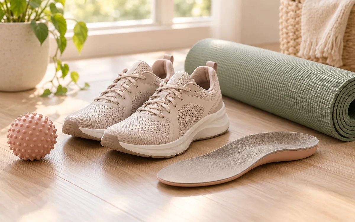
1. Fascia-Specific Stretching Exercises (The DiGiovanni Protocol): A large prospective randomized clinical trial published in The Journal of Bone and Joint Surgery by DiGiovanni and colleagues found that tissue-specific plantar fascia stretching yielded clearly better pain relief and functional restoration compared with conventional Achilles stretching alone [10]. To do this maneuver, sit comfortably and cross the affected foot over the other knee; grasp the base of the toes with your hand and gently dorsiflex them backward toward the shin until you palpate firm tension along the arch of your foot. Maintain this stretch for 10 seconds, repeating 10 repetitions before morning weight-bearing [2, 10]. 2. Gastrocnemius and Soleus Stretching: Stand facing a wall with the affected leg extended behind you, keeping the knee straight and heel firmly planted on the floor; lean forward until a strong stretch is felt throughout the posterior calf, holding for 30 seconds across three daily sets to improve ankle upward movement [1, 10]. 3. Footwear Optimization and Orthotics: Completely eliminate barefoot walking on unyielding household tile surfaces; instead, wear supportive indoor footwear featuring cushioned soles and structured arch contours. Select daily footwear with a firm heel counter and a modest heel-to-toe drop of 2 to 3 centimeters, which physically offloads the plantar fascia [1, 2, 4]. Prefabricated silicone heel cups or contoured arch-support insoles provide reliable shock dispersion and comfort [1, 6]. 4. Cryotherapy Massage: Rolling a frozen water bottle or firm massage ball beneath the foot arch for 10 to 15 minutes after workdays delivers effective topical analgesia and vasoconstrictive less inflammation [1, 2]. 5. Load Management and Low-Impact Conditioning: Temporarily substitute high-impact jogging with exercise that puts less weight on your foot, such as pool swimming or stationary cycling, to keep fitness while facilitating fascial tissue repair [2, 6].
If heel pain persists: treatment options
When heel pain persists past several weeks despite regular stretching and supportive insoles, physical medicine and rheumatology specialists provide advanced treatments to stimulate healing [1, 6]:
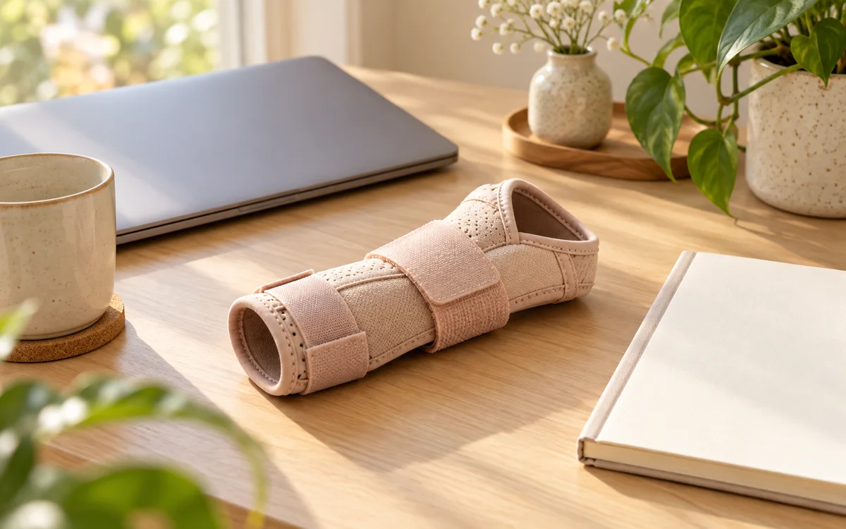
1. Extracorporeal Shockwave Therapy (ESWT): Radial shockwave therapy is an outstanding, non-invasive treatment treatment for chronic recalcitrant plantar fasciitis. A thorough meta-analysis by Guede-Rojas and colleagues found that targeted acoustic shockwaves promote localized new blood vessel growth, stimulate tissue repair, and induce immediate pain relief through sensory nerve desensitization, offering significant functional recovery without surgical exposure [13]. 2. Night Splints: Rigid or semi-rigid night splints keep the ankle and toe joints in neutral upward movement during sleep. By preventing the plantar fascia tightening overnight in a shortened position, night splints often alleviate the acute agony of initial morning weight-bearing [2, 6]. 3. Guided Corticosteroid Injections: While local corticosteroid delivery provides rapid short-term analgesia, a definitive Cochrane systematic review by David and colleagues demonstrates that its benefits rarely extend beyond four to twelve weeks. Clinicians must exercise stringent caution and use high-resolution ultrasound guidance, as unguided or repeated injections dramatically heighten the risk of catastrophic plantar fascia rupture and irreversible loss of the heel cushion [6, 14]. 4. Platelet-Rich Plasma (PRP) Injections: Autologous platelet concentrates provide concentrated growth factors that foster tissue repair, serving as an alternative biologic treatment for chronic tissue degeneration [6].
Surgical release of the plantar fascia remains an last option, reserved exclusively for the rare fraction of patients (fewer than 5%) who demonstrate persistent functional disability after at least 6 to 12 months of structured conservative therapy [2, 6].
Heel pain care at La Rose
If heel pain keeps getting in the way of your day, La Rose offers rheumatology care, joint and muscle ultrasound, and rehabilitation at two branches:
1. New Maadi Branch: 14 El-Mokhtar St, off El-Nasr St, behind QNB Bank, New Maadi. 2. Fifth Settlement Branch: CMC Medical Mall, 4th floor, North 90th Street, behind the Air Force Specialized Hospital.
Our rheumatology and rehabilitation medical team includes: - Dr Shaimaa Sherif: Physician and specialist in rheumatology, joint diseases, physical medicine, and rehabilitation; available at the New Maadi branch on Saturday, Monday, and Tuesday from 3 to 8 pm, and at the Fifth Settlement branch on Wednesday from 3:30 to 8 pm. - Dr Mohab Ashraf Fouad: Consultant in internal medicine, gastroenterology, and rheumatic conditions; available exclusively at the New Maadi branch on Saturday from 4 to 7 pm for complex multisystem reviews.
For specialist joint and entheseal evaluation in South Cairo, visit our dedicated rheumatology doctor in Maadi to structure physical therapy and medical management; for New Cairo residents, our specialized rheumatologist in Fifth Settlement offers assessment and rehabilitation care [3, 8]. Call or WhatsApp 010 4066 1893 to confirm your appointment.