What is gastroscopy and why is it recommended?
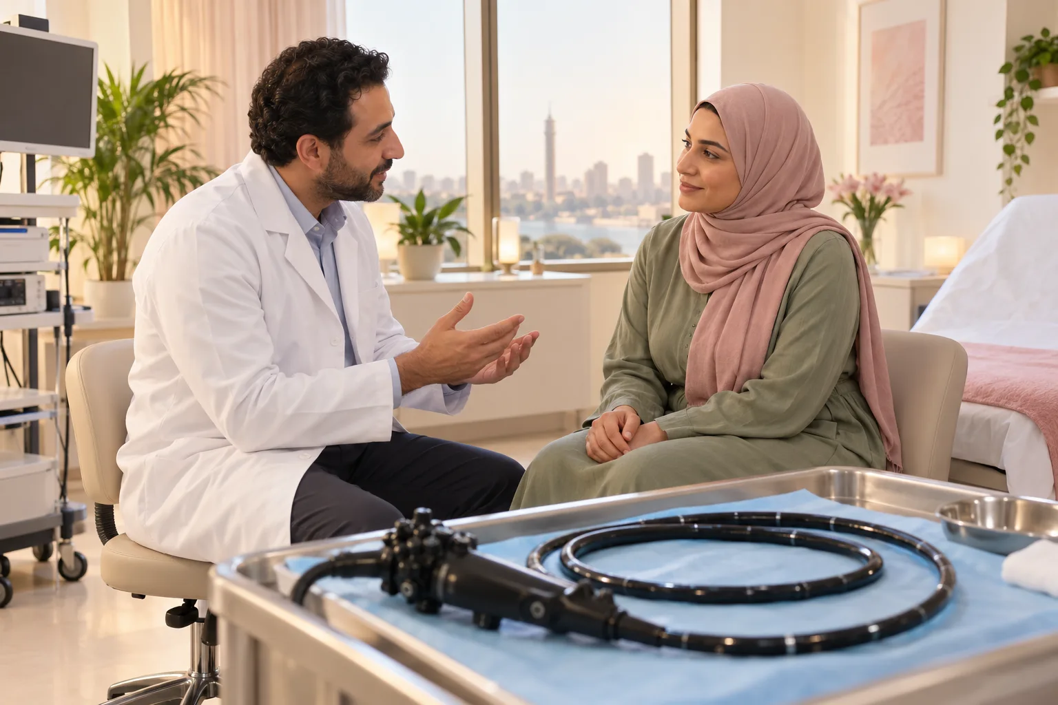
Gastroscopy is also called upper endoscopy or esophagogastroduodenoscopy (EGD). It is a precise, common test used to look closely at the inner lining of your upper digestive tract [1, 2, 3]. During the test, a doctor gently guides a thin, flexible tube through your mouth. This tube has a tiny high-definition camera and light at its tip. It passes down your food pipe (oesophagus), through your stomach, and into your duodenum, the first part of your small intestine [1, 2, 3]. The camera sends back clear, magnified pictures of your digestive lining in real time. This view is far more accurate than an abdominal ultrasound or a barium X-ray for spotting mild inflammation, surface scrapes, small peptic ulcers, or early changes in tissue [1, 3, 6, 7].
Your doctor usually recommends an upper endoscopy to answer a clear health question when your history and routine tests are not enough [1, 3, 4]. Common reasons include long-standing indigestion that does not improve with standard medicine, stubborn heartburn from acid reflux, unexplained upper belly discomfort, and lasting nausea or vomiting [1, 2, 4]. It is also very helpful for finding the cause of unexplained iron-deficiency anaemia, such as hidden blood loss or gut absorption problems like celiac disease. In addition, it helps your doctor check painful or difficult swallowing (dysphagia or odynophagia) [1, 4, 5, 9]. These goals are completely different from a lower bowel check, since upper endoscopy and colonoscopy look at entirely different areas of your body [1, 3].
International medical guidelines, including UK guidance from the National Institute for Health and Care Excellence (NICE CG184 and NG12), stress that gastroscopy is not a routine check for brief heartburn. Instead, it is a targeted test used when a physical illness must be ruled out, or when red-flag signs appear, such as unintended weight loss, repeated vomiting, or signs of bleeding [4, 5, 13]. Beyond examining the lining, the endoscope also acts as a channel for treatment. Through it, the specialist can take painless pinpoint tissue samples (biopsies) for lab checks, run quick germ tests, or treat problems directly, such as stopping bleeding or gently widening narrow areas [1, 3, 7, 13].
Preparation before gastroscopy: fasting and medication management
Careful preparation before your gastroscopy is vital to protect your safety and give your doctor a crystal-clear view of your digestive lining [1, 2, 3]. The most important basic rule is that your stomach and duodenum must be completely empty of solid food and cloudy liquids when the examination begins [1, 2]. Any leftover food or drink covers the lining of the stomach, preventing your examining doctor from spotting subtle raw patches, surface scrapes, or small ulcers. When this happens, the doctor often has to stop the procedure early and reschedule it for another day [1, 3]. Even more importantly for your personal safety, leftover food or liquid in your stomach while you are sedated greatly increases the danger of pulmonary aspiration, where stomach contents enter your lungs. Following standard fasting steps closely greatly lowers this risk [1, 2, 3].
Standard clinical prep requires you to stop eating all solid food for six to eight hours before your scheduled endoscopy appointment [1, 2, 3, 12]. You can usually drink modest sips of clear fluids (such as plain water or weak black tea without milk) up to two to four hours before the test, depending on your endoscopy unit's specific guidelines [1, 2, 12]. You must completely avoid milk, dairy drinks, cloudy juices, and carbonated beverages on the day of your procedure. You should also avoid smoking cigarettes and chewing gum, because both habits stimulate extra stomach acid and produce excess fluid inside the stomach cavity [1, 2, 3].
When it comes to your regular medicines, you must tell your doctor and nursing team about every prescription pill, over-the-counter remedy, and herbal supplement you take well ahead of time [1, 2, 3]. Blood-thinning and antiplatelet medicines (such as warfarin, clopidogrel, aspirin, or direct oral anticoagulants) require individualized instructions from your doctor on whether to pause or adjust them, especially if tissue samples or treatments are planned. You should never pause or stop these medicines on your own [1, 3]. You can usually take essential heart and blood pressure pills at your normal morning time with a tiny sip of water to keep your blood pressure steady. In contrast, diabetes tablets and insulin doses are carefully adjusted to match your fasting and prevent dangerous drops in blood sugar [1, 2, 3]. In addition, if your doctor plans to take biopsies to check for Helicobacter pylori bacteria, you may need to pause proton pump inhibitors (PPIs) for a set time so the test gives an accurate result [10, 11].
How gastroscopy is performed: a step-by-step procedural walkthrough
Many people want to know what happens step by step on the day of a gastroscopy inside the procedure room [1, 2, 3]. Once standard safety checks are finished and your baseline vital signs (blood pressure, heart rate, and oxygen levels) are recorded, the nursing team helps you lie down comfortably on your left side on the examination bed. This left-sided posture aligns your digestive tract naturally and helps saliva drain easily without causing coughing [1, 2, 3]. Any removable dentures, dental plates, or eyeglasses are safely set aside beforehand to protect them and give the clinical team clear access to your mouth [1, 2, 12].
A small, smooth plastic mouthguard is placed gently between your teeth and secured with a soft elastic strap behind your head. This protects your teeth and keeps you from accidentally biting down on the delicate camera tube [1, 2, 3]. The clinical team then sprays a local numbing medicine onto the back of your throat, or gives you mild intravenous sedation through a small cannula in your arm, depending on your agreed plan [1, 2, 3]. The doctor puts a smooth lubricant on the tip of the slender, flexible tube and guides it over your tongue toward the back of your throat. When you gently swallow, your food pipe relaxes open, allowing the scope to slide smoothly into your oesophagus without causing injury [1, 2, 3].
Throughout the whole test, your normal breathing is completely open and unobstructed. The tube travels down your food pipe (oesophagus), leaving your voice box, windpipe, and airway entirely free so you can breathe steadily and calmly through your nose and mouth [1, 2, 3, 12]. To get a complete, clear view of the digestive lining, the doctor gently introduces small pulses of air or carbon dioxide through a tiny channel inside the scope. This gently opens up the natural folds of your stomach and duodenum, giving a detailed high-definition view of the entire surface [1, 2, 3]. The specialist systematically checks the lining of your food pipe for reflux irritation, small breaks, or swollen veins, inspects every part of your stomach, and moves into the first two sections of your duodenum [1, 3, 6, 7]. A standard diagnostic upper endoscopy usually takes between five and fifteen minutes of active scope time. Once finished, the doctor suctions out the extra air to relieve fullness, then withdraws the tube gently and swiftly [1, 2, 3].
Is gastroscopy painful? Sedation and local anaesthesia options
The most common concern people have is whether a gastroscopy hurts and whether being put completely to sleep is necessary [1, 2, 3]. The medical answer is reassuring: gastroscopy does not cause sharp, cutting pain because the inner lining of your digestive tract does not have the sharp pain receptors found in your skin [1, 2, 3]. If you have the test without sedation, what you feel is not sharp pain, but rather mild pressure in your throat and upper stomach, along with a quick, natural gagging reflex as the tube passes the back of your throat. Afterward, you may feel temporary abdominal fullness from the air used during the examination [1, 2, 3, 12].
You and your doctor can choose between two main options to ensure your comfort, depending on your health and personal preference [1, 2, 3]. The first option is a numbing throat spray, usually lidocaine, applied to the back of your tongue and throat walls [1, 2, 3]. Within sixty seconds, this spray numbs your throat lining, which greatly softens your gag reflex and helps the scope glide down smoothly [1, 2, 3]. With numbing spray alone, you stay fully awake, clear-headed, and able to follow simple requests. The big advantage is a very fast recovery, allowing you to walk out of the clinic on your own right away without needing an escort or taking time off from your normal day [1, 2, 12].
The second option is mild intravenous conscious sedation, where fast-acting relaxing medicines are given through a small cannula in your vein to help you feel deeply calm, drowsy, and free from worry [1, 2, 3]. While sedated, you breathe on your own and can still respond to gentle spoken words, but you feel very little awareness and usually remember nothing about the test afterward [1, 2, 3]. In certain clinical situations, deeper sedation with propofol given by an anaesthetist provides complete unconsciousness during the short examination [1, 3]. Importantly, medical practice varies by facility, reason for the test, and personal choice: not every gastroscopy requires sedation, nor is every patient a suitable candidate for sedatives, so your doctor will tailor the comfort plan directly to you [1, 2, 3].
Gastric biopsy: why tissue samples are taken and what they mean
People often feel quite anxious when they hear that small tissue samples (biopsies) were taken during their gastroscopy, often jumping to the frightening conclusion that the doctor suspects cancer or a life-threatening illness [1, 3, 12]. In clinical reality, taking small tissue samples is a standard, routine, and everyday part of diagnostic endoscopy. In the vast majority of cases, it is done to evaluate harmless, treatable stomach conditions that cannot be fully identified just by looking through the camera [1, 3, 6, 12]. Using tiny biopsy forceps passed through a channel inside the flexible tube, the specialist gently pinches off tiny specks of the lining (measuring merely one to two millimetres) without causing any pain or physical discomfort [1, 2, 3].
There are many harmless and routine medical reasons for taking tissue samples during an upper endoscopy [1, 3, 6]. One of the most common reasons is testing for Helicobacter pylori, a bacterial infection that can be checked right away in the clinic with a quick test or examined under a microscope to choose the right antibiotic treatment [1, 6, 11]. Biopsies are equally important for assessing the type, depth, and stage of chronic stomach inflammation (gastritis), checking for thinned tissue or chemical irritation [6, 11]. In addition, taking tissue samples from the first parts of your small intestine (the duodenum) is the gold standard for checking celiac disease, confirming microscopic changes in the gut lining even when the surface looks completely normal to the naked eye [9, 10].
Taking tissue samples is also routine in people with long-standing acid reflux to check for and monitor Barrett's oesophagus, and to confirm that stomach ulcers are non-cancerous before beginning healing medication [4, 7, 8]. You can feel reassured that sampling tissue is a careful, routine diagnostic method that removes guesswork and confirms an exact diagnosis [1, 3, 6]. The tissue samples are placed in preservative and sent to an accredited pathology laboratory, with detailed microscopic reports usually ready within several days to guide your ongoing medical care [1, 3, 12].
Recovery and aftercare: returning to eating and post-procedure rest
After your endoscopy is complete, you are moved to the recovery area for regular monitoring. You typically rest under nursing care for thirty to sixty minutes until your vital signs are steady and any sedative medicine wears off [1, 2, 3]. If you received only the numbing throat spray, you stay fully awake and can sit up right away. In contrast, if you received intravenous sedation, you will need a quiet recovery period under observation to regain full mental alertness and steady balance before you start walking [1, 2, 12].
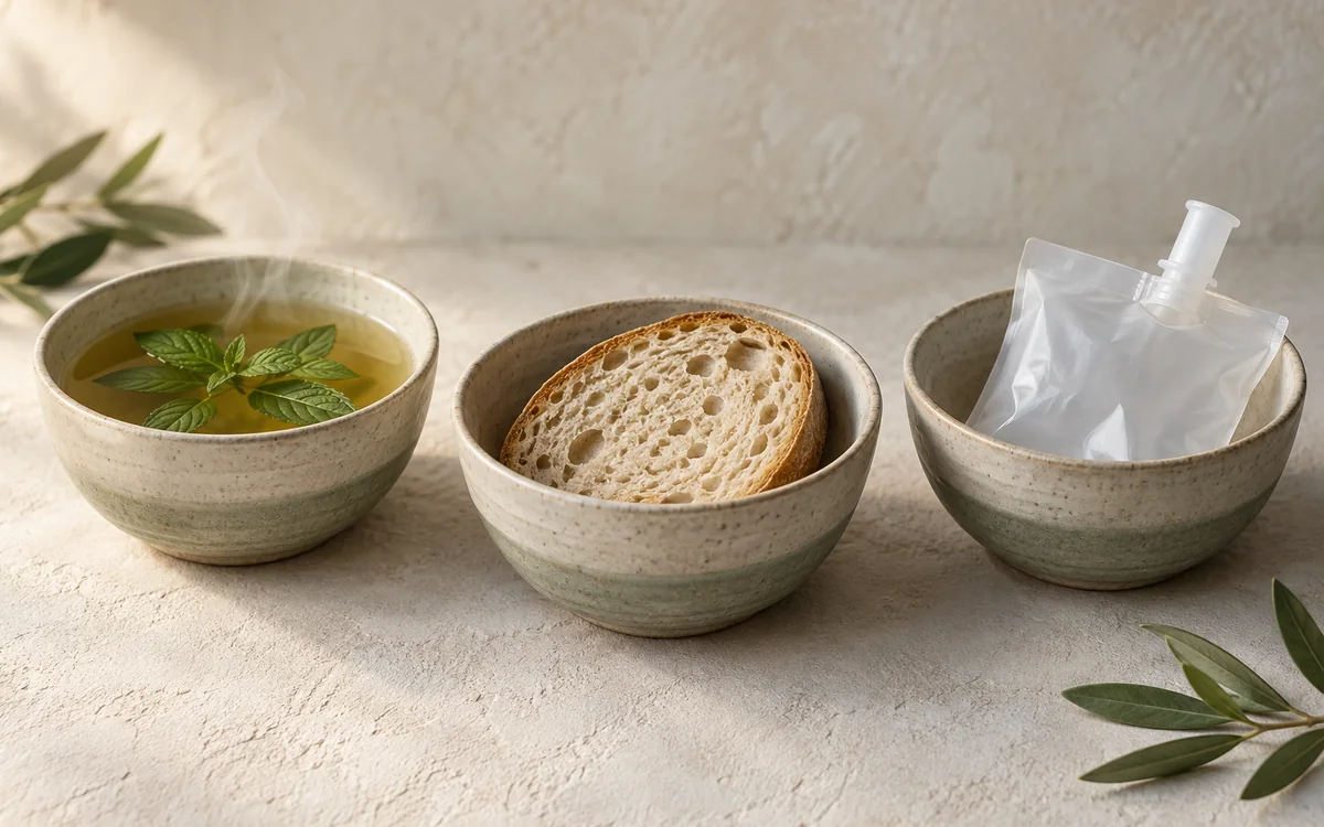
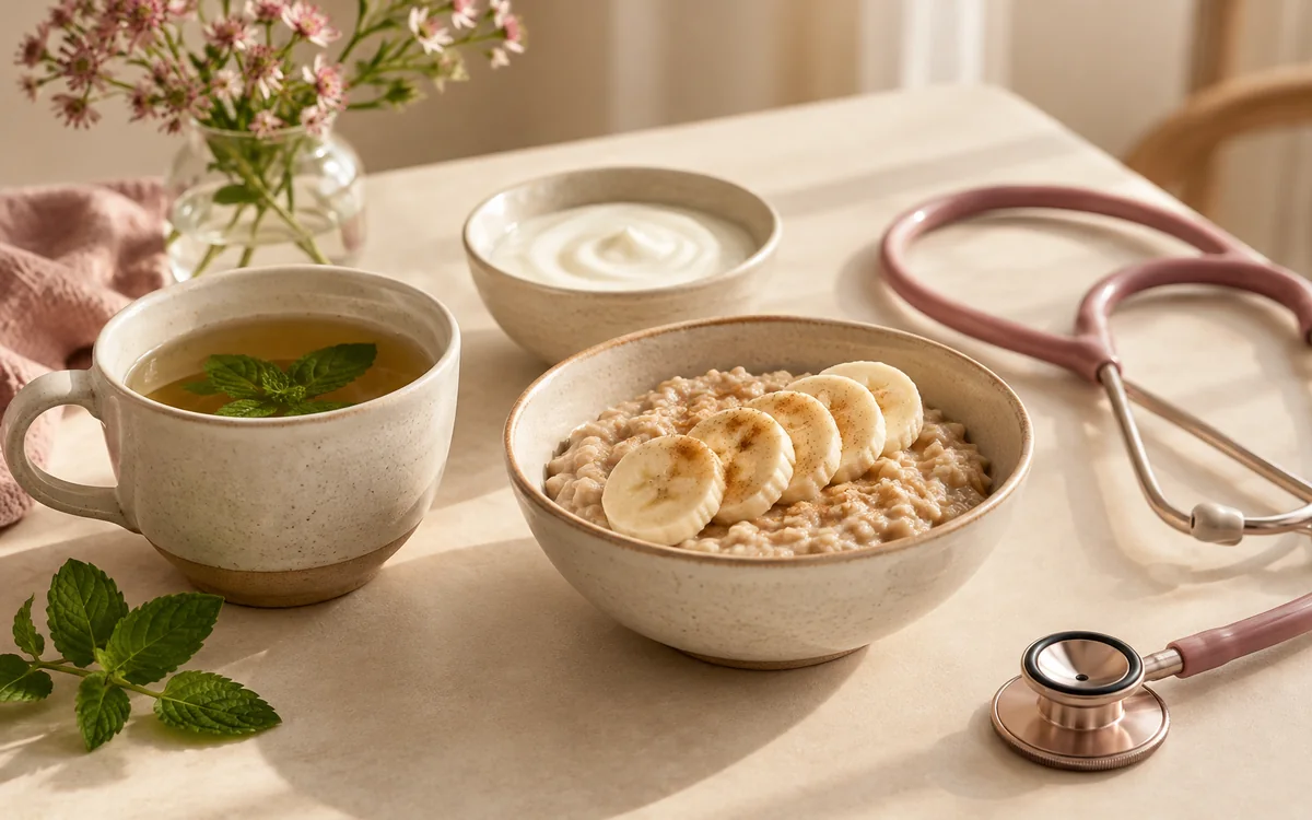
Knowing when you can eat and drink again is a common question, and the main rule depends on waiting for the numbing throat spray to wear off completely [1, 2, 3]. The numbness in your throat generally lasts for forty-five to sixty minutes after it is sprayed. During this time, you must not eat or drink anything, because your normal swallowing reflex is numbed and you could choke or accidentally breathe fluids into your lungs [1, 2, 12]. Once normal feeling returns and your throat feels clear, you can take a cautious test sip of plain water to ensure you can swallow easily [1, 2, 3]. After that, you can enjoy a light, gentle meal such as warm broth, yogurt, or soft bread, while heavy, spicy, or greasy foods and fizzy drinks should be avoided for the rest of the day to keep your stomach settled [1, 2, 3].
It is completely normal to feel mildly bloated, experience gentle cramps, or feel an urge to burp during the first few hours after your test. This is just temporary gas left over from the air used during the examination, which clears quickly through passing wind and gentle walking [1, 2, 3]. A slightly dry or scratchy throat, similar to the early start of a common cold, is also common and typically fades away within twenty-four to forty-eight hours [1, 2, 12]. Most importantly, if you had intravenous sedation, you must follow strict safety rules: an adult companion must take you home, and you must avoid driving a car, operating complex machinery, or signing important legal or financial papers for a full twenty-four hours until your mind is completely clear [1, 2, 3, 12].
Post-procedure warning signs and follow-up with the medical team at La Rose
Gastroscopy is recognized internationally as one of the safest and most reliable diagnostic tests in gastroenterology, with major complications occurring exceptionally rarely when performed by experienced endoscopists [1, 2, 3]. Nonetheless, patient safety requires clear knowledge regarding clinical red flags that necessitate immediate contact with the endoscopy unit or prompt presentation to an emergency department [1, 2, 3, 12]. It is clinically essential to differentiate between benign, temporary sensations (such as mild throat soreness or passing gas) and acute complications requiring swift intervention [1, 2, 12].
Warning signs that need immediate medical help include severe or worsening pain in your chest, neck, or belly that does not ease with routine care, or growing trouble breathing or swallowing [1, 2, 3, 12]. You also need urgent medical evaluation for lasting fevers, shaking chills, or nonstop vomiting that prevents you from keeping fluids down [1, 2, 12]. In addition, signs of bleeding in your upper digestive tract require emergency care right away. These include vomiting bright red blood, vomiting dark material like coffee grounds, passing pitch-black tarry stools (melena), or feeling faint, dizzy, and unsteady upon standing [1, 2, 8, 12, 13]. If you notice any of these symptoms after your endoscopy, get emergency medical care without delay [1, 2, 3].
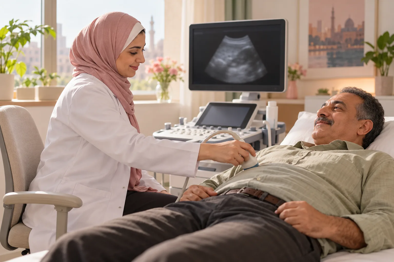
At La Rose Clinics, the medical team provides structured, careful clinical evaluations for individuals experiencing chronic upper gastrointestinal symptoms or who may need an endoscopy. Your care pathway begins with a thorough medical consultation, physical examination, and selective non-invasive tests (such as an in-clinic abdominal ultrasound or targeted blood work) to determine whether an upper endoscopy is genuinely needed. When a gastroscopy is recommended, the medical team ensures strict procedural safety and tailored patient comfort, followed by a clear, supportive explanation of what the doctor found and any biopsy results without confusing medical jargon or unrealistic promises. When you experience persistent upper digestive discomfort and require structured clinical clarity, you can book a consultation with the medical team to initiate your comprehensive evaluation.