Why the abdomen changes after pregnancy
During pregnancy the womb grows and pushes the abdominal wall forwards, and the muscles and the connective tissue that holds them together stretch to make room for the baby. This change is normal, and most of it recovers gradually over the months after birth. [7]
The extra pressure on the abdominal wall can, however, reveal a weak spot, especially around the belly button, which is why pregnancy is a recognised factor that raises the chance of a hernia. A caesarean birth also adds an incision in the abdominal wall, and a scar can later become the site of an incisional hernia. [4, 10]
So a bulge after birth has more than one possible explanation: separated abdominal muscles, an umbilical hernia, a hernia at the caesarean scar, or a groin hernia. Each looks different and each has a different plan. [2, 7, 10]
This article helps you tell these possibilities apart in a first, general way, and explains when a bulge deserves a routine examination and when it needs emergency care. A definite answer, however, comes from an examination. [2, 7]
Separated abdominal muscles: the most common explanation
Rectus abdominis separation means the two muscles running down either side of the middle of the abdomen move apart, and the tissue between them becomes thinner and weaker. It is very common after pregnancy and shows as a long ridge or dome down the middle of the abdomen when you sit up from lying down or tense your abdominal muscles. [7]
The important difference is that separation is not a hole in the wall, and no bowel pushes through it and becomes trapped. So it is usually not dangerous, even if it looks bothersome or affects how strong the abdomen and back feel. [7, 10]
Much of this separation improves on its own in the weeks and months after birth, and exercises set by a physiotherapist help strengthen the deep abdominal muscles. North Bristol NHS Trust advises avoiding exercises that put extra pressure on the middle of the abdomen at first, such as traditional sit-ups, until an assessment has been done. [7]
If the separation is still obvious after the recovery period, or if there is pain or a bulge at one specific point, the examination looks for an umbilical or epigastric hernia alongside it, because the two can occur together. [7, 8]
Umbilical hernia after pregnancy
An umbilical hernia shows as a soft bulge inside or around the belly button and can make the belly button stick out. It gets bigger when you cough, stand or strain and smaller when you lie on your back. [2, 4]
Pregnancy is one of the commonest reasons an umbilical hernia appears in adults, because pressure inside the abdomen is raised for months. Sometimes the hernia shows during the pregnancy itself and sometimes after the birth. [4, 10]
Unlike muscle separation, an umbilical hernia sits at one specific point, and the doctor may feel a small hole in the wall beneath the bulge. If the hole is small, the bulge can hurt with effort more than its size would suggest. [2, 10]
In adults an umbilical hernia does not close on its own, as it sometimes does in children. That does not mean surgery must happen straight away; timing is decided after an examination according to size, symptoms and plans for future pregnancies. [2, 8]
In the meantime, things that repeatedly raise pressure inside the abdomen, such as an untreated chronic cough, constipation with heavy straining, or lifting heavy loads without support, can make the bulge more obvious or more uncomfortable. Treating constipation and cough, and lifting children and heavy items in a way that supports the abdomen, are simple steps that make daily life more comfortable until a decision is made. Weight gained during pregnancy also adds pressure on the wall, so a gradual return to a healthy weight helps, without harsh dieting while breastfeeding. [1, 4]
A bulge at the caesarean scar
Any operation that opens the abdominal wall, including a caesarean section, can be followed by an incisional hernia at the scar. It shows as a bulge along the scar or at one end of it and can appear months or even years after the operation. [8, 10]
Some factors raise the chance of an incisional hernia, such as wound infection after the operation, excess weight, smoking, a chronic cough and diabetes. Having one of them does not mean a hernia will definitely happen, but it makes it more important to notice any new bulge. [8, 10]
Not every swelling above a caesarean scar is a hernia. Sometimes the skin and fat above the scar form a fixed overhanging fold that does not change when you cough, and that is not a hernia. A hernia usually grows with pressure, and the doctor may feel a hole in the wall beneath it. [2, 10]
CT or ultrasound may be requested for an incisional hernia to show the size of the defect and whether there is more than one, which matters for planning if repair is chosen. [8, 10]
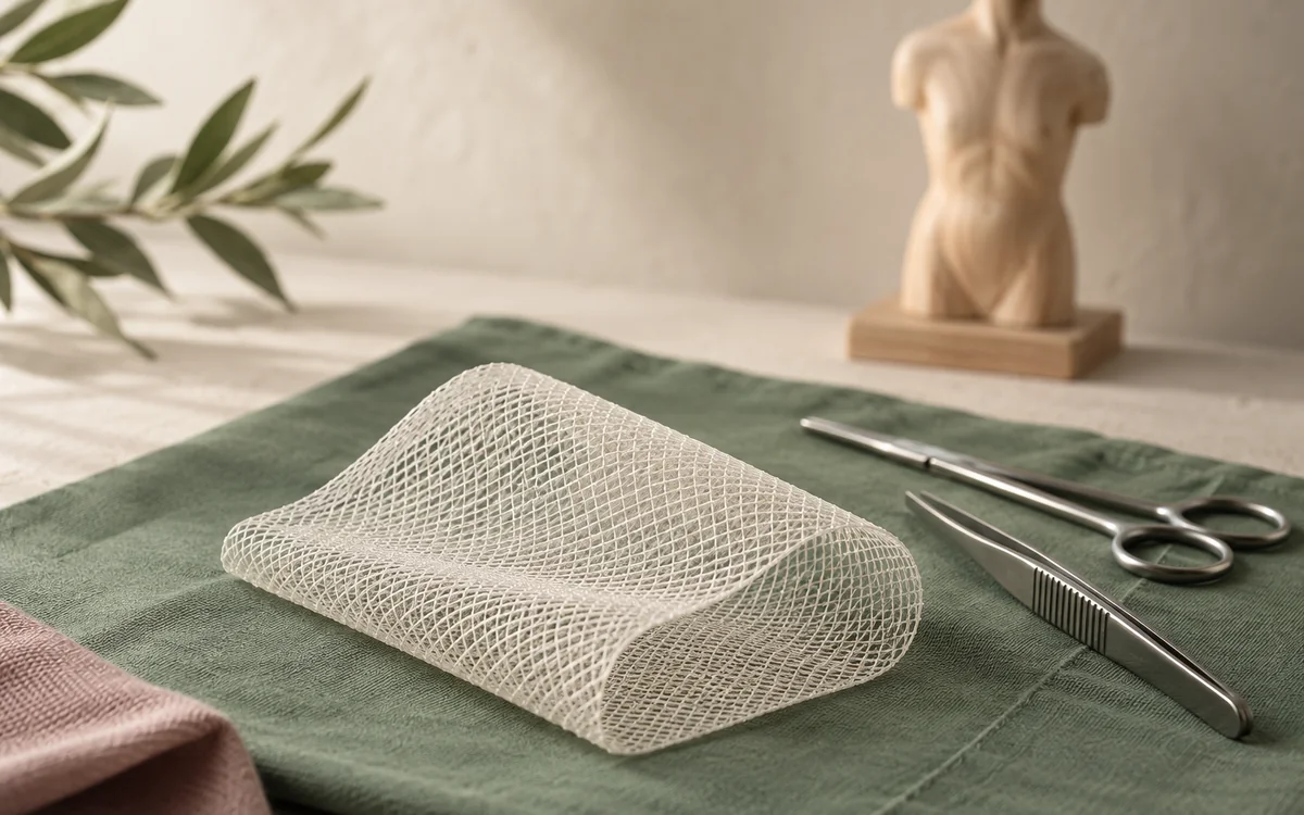
An incisional hernia can be repaired by open surgery or keyhole surgery, and mesh is often used to reinforce the wall. The choice of approach depends on the size and site of the defect, previous operations and general health, and it is discussed in detail with the surgeon before any decision. [8]
C-section hernia symptoms: compare the scar, midline and belly button
A C-section scar hernia is an incisional hernia at a previous surgical cut. The usual clue is a visible bulge on or near the scar, especially when coughing or straining, sometimes with pain or pressure at the same site. An umbilical hernia is centred around the belly button, while diastasis recti after pregnancy is widening between the abdominal muscles in the midline rather than one focal scar lump [4, 7, 11].
This comparison can help you describe the change, but it cannot confirm the diagnosis at home. Arrange an examination for a new C-section scar bulge or persistent pain or pressure. Seek urgent care for sharp abdominal pain with vomiting, or when the bulge is painful with bloating, constipation or fever; the American College of Surgeons identifies sharp pain with vomiting as a possible strangulated bowel in the hernia and a surgical emergency [2, 11].
Groin and femoral hernias in women
After childbirth a bulge or pain can appear in the groin or at the top of the thigh. Inguinal hernias are less common in women than in men, but femoral hernias make up a larger share in women, and this type is more likely to become trapped. [2, 6]
A femoral hernia appears below the groin crease at the top of the thigh and is often small. In women a hernia in this area can also show as pain without an obvious bulge, so the doctor needs to look for it specifically. [6]
The HerniaSurge guidelines advise that women with a groin hernia are offered repair rather than watchful waiting, because telling an inguinal from a femoral hernia on examination is not easy. Ultrasound helps when symptoms are present but the examination is unclear. [6]
So any new bulge in the groin or upper thigh after childbirth deserves a prompt examination, and any bulge there that becomes firm and painful needs emergency care. [2, 6]
When a bulge needs emergency care
Muscle separation does not become trapped, but a hernia can. The signs that need emergency care straight away are sudden severe pain at the bulge, a bulge that has become firm and painful and does not go back when you lie down, or redness or a change in the colour of the skin over it. [2, 4]
With a bulge, vomiting, being unable to pass wind or open your bowels, or fever are signs that the bowel may be affected, and that cannot wait for a clinic appointment. [2, 5]
After a caesarean in particular, some symptoms need prompt contact with the maternity team even without a hernia: redness of the wound or discharge from it, fever, or pain that is getting worse instead of better. These can be signs of wound infection, which is also one of the factors that raises the chance of a hernia later. [8, 10]
The full list of warning signs is in our article on abdominal hernia warning signs, and it is useful for the whole family to know. [2]
How the examination tells them apart
The doctor starts with the story: when the bulge appeared, exactly where it is, whether it changes with coughing and lying down, whether there is pain, whether the birth was vaginal or by caesarean, and whether the wound became infected afterwards. [2, 4]
Next comes an examination of the abdomen while you stand and lie down, with a request to cough or lift your head from the pillow. With muscle separation the bulge shows as a long ridge in the middle; with a hernia it sits at one point and a hole may be felt beneath it. [7, 10]
A scan is not needed in every case. Ultrasound helps when the examination is unclear or there is pain without a bulge, and CT helps with a large incisional hernia or when more than one defect is suspected. [6, 8, 10]
If you would like more detail on how symptoms look with each type of hernia, you can read the abdominal-wall hernia symptoms guide before the appointment. [2, 3]
The plan after diagnosis
If the diagnosis is muscle separation, the plan is usually physiotherapy and graded exercises, with the improvement followed up over the months. Surgery is not the first choice in that situation, and improvement depends on patience and regular exercise more than anything else. [7]
If the diagnosis is a hernia, the surgeon discusses whether repair is needed now or whether it can be watched, and which type of operation fits. The things that enter the decision are the size, symptoms, type of hernia, general health, plans for future pregnancies, smoking and weight. [6, 8]
A belt or binder may make some people more comfortable, but it does not treat the hernia or close the hole, and it does not replace an examination. Forcing a bulge back in is not safe if it is painful or firm. [2, 4]
Before the appointment, write down where the bulge is, when it appeared, the type of birth, any wound problems and your medicines. If there is a bulge after birth that has not been examined, or a known hernia that is changing, you can book an appointment for an examination and a plan. [1, 8]