
When Do You Need an Abdominal Ultrasound? The Direct Answer
You need an abdominal ultrasound when the clinician has a focused question that a picture may answer: are gallstones causing pain under the right ribs, does the liver look fatty, are the kidneys swollen from blocked urine, or is there fluid in the abdomen? It is not a random test for every episode of wind or pain, but a useful next step when symptoms, examination or blood tests point to an abdominal question [3, 7].
The scan can assess the liver, gallbladder, bile ducts, kidneys, spleen, pancreas and major abdominal blood vessels to differing degrees, and it uses sound waves rather than X-rays [3, 5]. That makes it a sensible first imaging test for selected upper-abdominal pain, jaundice, abnormal liver or kidney tests, suspected stones, organ enlargement, or follow-up of a known finding [3, 6].
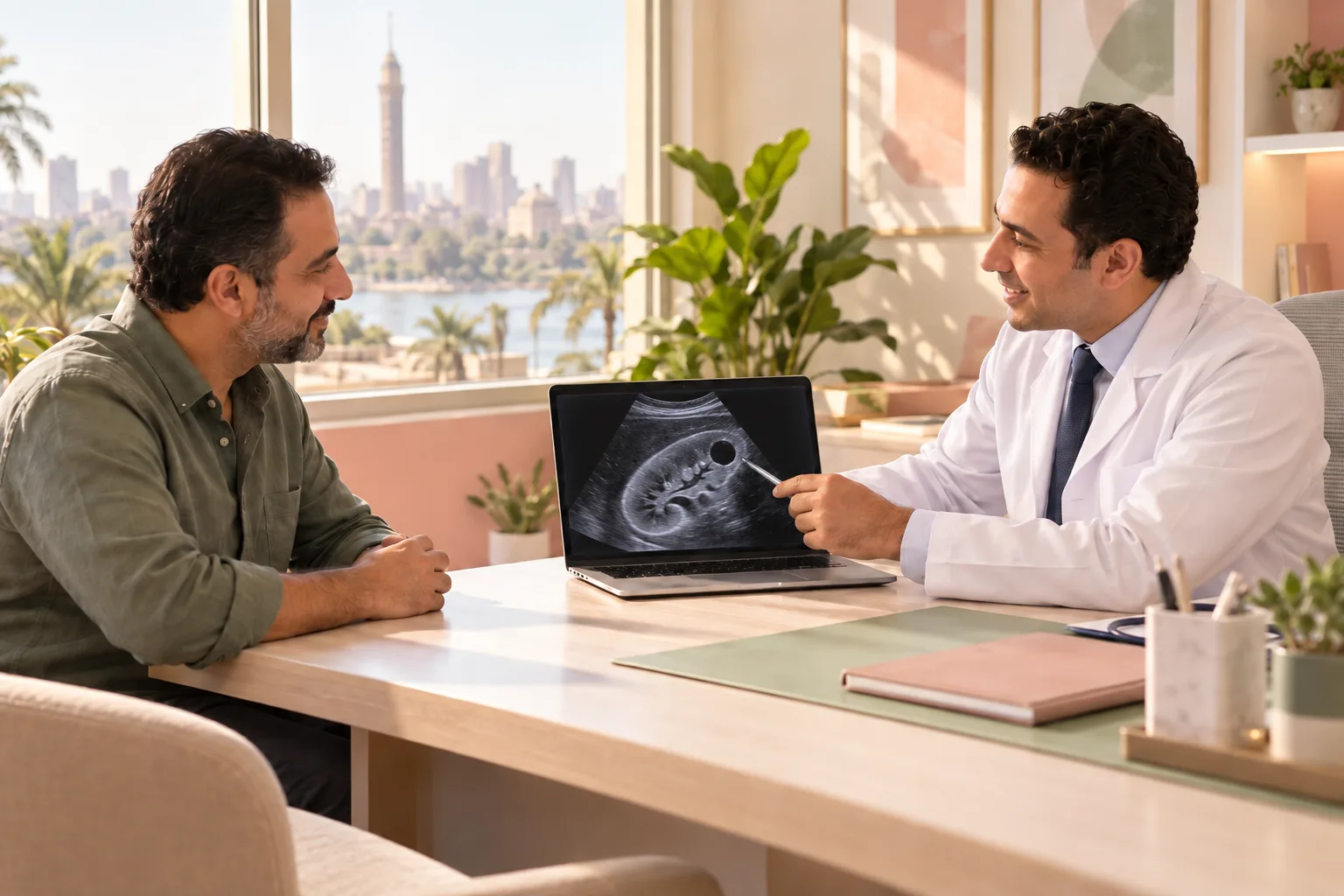
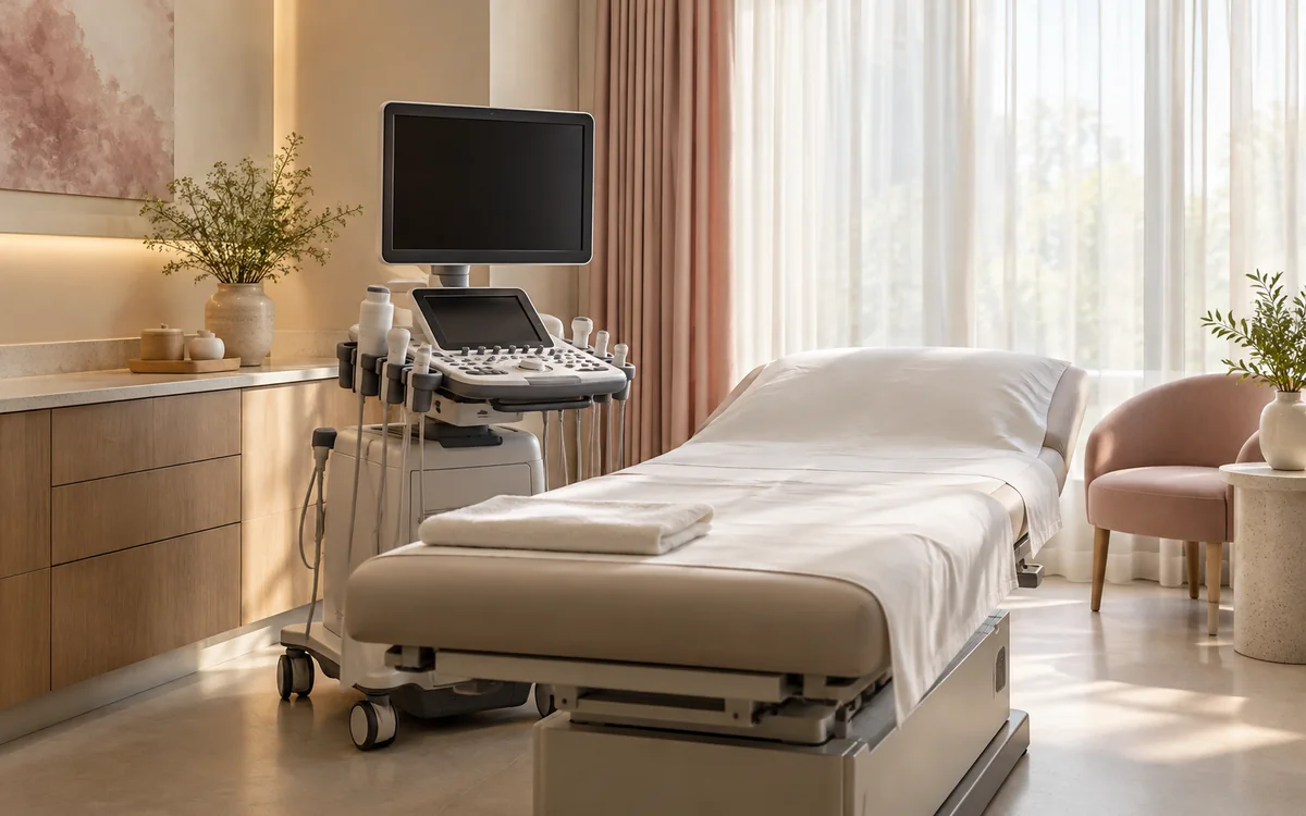
At La Rose Clinics in Maadi, Dr Mohab Ashraf Fouad, a specialist in internal medicine, diabetes, liver disease, gastroenterology and endoscopy, can perform an abdominal ultrasound in the same visit when it suits the clinical question. He is available on Saturday from 4 to 7 pm at 14 El-Mokhtar St, off El-Nasr St, behind QNB Bank, New Maadi. Book your appointment; schedules are confirmed when booking and can change.
From Symptom to Question: What Is the Scan Looking For?
The location and pattern of pain matter more than the vague phrase “stomach ache”. Steady or repeated pain beneath the right ribs, especially after a rich meal and with nausea, raises a practical question about the gallbladder, bile ducts and stones. A stone can be found incidentally without causing symptoms, so a report finding does not automatically explain every episode of bloating or mean treatment must begin immediately [9].
Pain in the side or lower back that travels towards the groin, with urinary burning or a change in urine, may prompt a scan for kidney swelling or a visible stone. In severe adult pain, however, urine and blood tests or different imaging may also be needed because ultrasound alone does not always settle the location of every stone [13].
Upper-abdominal heaviness, yellow skin or eyes, itch with dark urine, or abnormal liver blood tests can create a question about the liver, gallbladder, ducts and fluid around organs. If the abdomen enlarges quickly or the clinician feels a lump or enlargement, ultrasound can help distinguish fluid, an enlarged organ or a finding needing more detailed imaging [3, 5].
Here is an everyday contrast. Someone gets right-sided upper pain after fried breakfast twice a month, so the useful question is, “Are there gallstones or signs of gallbladder inflammation?” Someone else has long-term bloating and constipation but a normal examination and blood work; ultrasound may reassure about selected organs, but it cannot diagnose irritable bowel syndrome by itself [8, 12].
What Can an Abdominal Ultrasound Actually Show?
Ultrasound is particularly useful for questions involving visible organs and fluid: gallstones, widened bile ducts, liver appearance, kidney cysts or swelling, spleen size, abdominal fluid, and sometimes measurement of the abdominal aorta. If Doppler is requested, it adds a question about the direction and speed of blood flow in specified vessels [5].
For fatty liver, a routine ultrasound may show fat in the liver, which is one reason to request it with higher body weight, diabetes, raised triglycerides or abnormal liver enzymes. A fat appearance alone cannot tell whether there is inflammation or scarring, so the next step may include blood tests, risk scores or a liver-stiffness test according to the whole clinical picture [10]. Read What abdominal ultrasound shows to separate a written finding from what it means in your own situation.
If a report says “simple kidney cyst”, that is an imaging description to interpret with age, symptoms and the rest of the report, not a diagnosis to fear or dismiss without discussion. The same principle applies to words such as “enlarged” or “fatty”: a measurement or appearance starts a clinical question; it does not close the story [3, 10].
What an Ultrasound Cannot Settle
An ultrasound is not a camera that sees everything in the abdomen. Gas and air in the bowel weaken the image, so the ordinary scan has limited ability to examine the intestines, stomach and colon. A thicker abdominal wall or recent barium contrast from another test can also reduce the clarity of some organs [1, 8].
That means a normal scan does not rule out every cause of pain. If the question is about the stomach lining or an ulcer, endoscopy or specific tests may fit better. If pain is acute or the image does not answer the question, the clinician may choose CT, MRI or close review instead of repeating the same scan without purpose [4, 5]. The useful post-report question is, “Did the scan answer the reason it was requested, or is there still a different question?”
One important example is that ultrasound does not confirm or exclude colon cancer because it cannot reliably view the colon lining through bowel gas. Can an abdominal ultrasound detect colon cancer explains why symptoms and risk factors determine whether a different bowel test is needed. If the report is normal but pain persists, A normal ultrasound does not end assessment explains when to revisit the plan rather than treating the result as a final verdict.
Fasting and Preparation: What to Do Without Guessing
Do not assume every abdominal ultrasound has identical preparation. Many examinations require fasting for several hours, and sometimes 8 to 12 hours, because food and bowel gas can block the view of the gallbladder or weaken the image. The instruction from the imaging team and the exact type of scan are the deciding factors, particularly if the request includes the pelvis, kidneys or bladder [2, 4, 6].
Take regular medicines only as instructed. Medical sources note that a small sip of water with medicine may be acceptable in some arrangements, but a person with diabetes or medicine linked to meals needs to ask before fasting so the plan can be adjusted safely [1, 8]. Do not stop a medicine on your own simply because you have an ultrasound.
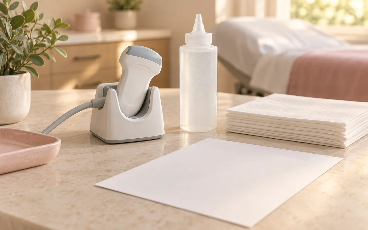
On the day, wear comfortable clothing and bring the request, previous reports, recent blood results, and photographs of medicine boxes or a clear medicines list. You will lie on your back or side; gel goes on the skin, the probe moves over the abdomen, and you may be asked to hold your breath briefly or change position. The examination often takes under 30 minutes, and pressure over a sore spot may be uncomfortable but should not create new pain [1, 3]. Ultrasound preparation card brings together the fasting, water and medicine questions to check before the appointment.
After the Result: Three Common Scenarios
A result does not divide neatly into only “good” or “bad”. Scenario one: the report finds gallstones and your symptoms really are typical right-rib pain. The clinician then matches the picture with pain duration, vomiting, fever and blood tests, because silent gallstones are common and not every stone calls for the same action [9].
Scenario two: the report says fatty liver. That is a reason to review blood pressure, glucose, blood fats, medicines, food pattern and movement, not a shortcut to judging the degree of liver damage. Routine imaging can show fat but cannot establish inflammation or fibrosis, so blood work and a further risk assessment may be appropriate according to individual risk [10]. To understand what the scan clarifies and what requires additional assessment, read our guide on fatty liver ultrasound before your follow-up visit.
Scenario three: “no clear cause on ultrasound” with ongoing pain. This is not an empty result. It reduces the likelihood of some problems in the organs that were visualised, but it does not erase stomach, bowel or non-abdominal causes. The clinician returns to the symptom map: location, relationship to food or urination, weight loss, bowel change, examination and tests, then chooses review or a different test [4, 8].
Keep the written report rather than only a mobile photo. Mark the “impression” or conclusion and note why the scan was requested, the symptoms that day, and whether there is an older scan for comparison. Those details make the next conversation more precise than asking only, “Was it normal?”
When Not to Wait for a Scan Appointment
Ultrasound should support assessment, not delay emergency care. Go to emergency care now if abdominal pain comes on suddenly and is severe, the abdomen is very painful to touch, there is vomit containing blood, black or bloody stool, collapse, breathing difficulty, chest pain, or inability to pass urine, stool or wind [12].
Pain that began around the navel and within hours moved to the lower right abdomen, worsening with movement or coughing, needs urgent assessment for possibilities including appendicitis. Examination, ultrasound or other imaging, and blood tests are selected in the appropriate setting; a routine clinic scan is not a substitute for emergency assessment when symptoms are accelerating [11].
Severe pain under the right ribs or in the middle of the abdomen with vomiting, fever or shivering, or yellow skin and eyes also needs urgent care. This can occur with gallbladder-related blockage or inflammation, when speed matters more than waiting for a routine result [9]. Severe side or back pain with fever or shivering, particularly with cloudy urine or vomiting, likewise needs urgent assessment for a possible stone or urinary infection [13].
If pain is tolerable but persists for days, returns repeatedly, wakes you at night, or comes with unintentional weight loss or persistent bloating, arrange a prompt review. The point is not to worry about every symptom; it is to match the timing and imaging to the pattern rather than to a hoped-for report.
Abdominal Ultrasound in Maadi: Making the Visit Useful
If you are looking for an internal-medicine doctor who can examine with ultrasound in Maadi, prepare the visit around a clear question rather than a bare request for a scan. Before arriving, note where the pain is, when it began, whether it follows a particular food or urination, and whether there is fever, vomiting or yellowing. Bring old blood tests and reports. This helps Dr Mohab decide whether an abdominal ultrasound in the same visit answers the right question and what needs to be imaged.
Dr Mohab Ashraf Fouad sees internal medicine, diabetes, liver and gastroenterology cases and can perform abdominal ultrasound in the same visit when needed, on Saturday from 4 to 7 pm at 14 El-Mokhtar St, off El-Nasr St, behind QNB Bank, New Maadi. To book, call or send a WhatsApp message to 010 4066 1893; schedules are confirmed when booking and can change. Abdominal ultrasound provides a safe, real-time evaluation of internal abdominal organs using sound waves without any radiation exposure [1].
After the result, leave with a short written plan: does it explain the symptom, is a blood test, follow-up or a different specialty needed, and which warning sign means do not wait? Abdominal ultrasound in Maadi explains the visit pathway, and Internal medicine doctor in Maadi helps you organise questions when several symptoms or blood results need to be considered together.
If the examination cost is on your mind, send a WhatsApp message for the details rather than relying on advertisements or old figures. Mention whether the request is for abdomen only or includes another examination, because the scan type and preparation change what is needed before the visit.