The device measures impedance, then estimates composition
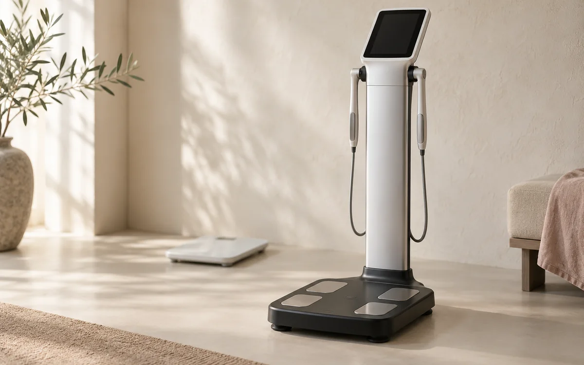
InBody is a form of bioelectrical impedance analysis, or BIA. The device passes a very small electrical current between electrodes and measures tissue resistance and other electrical properties. Water-rich tissues conduct current differently from fat. It then combines the measurement with height, weight, age or other inputs in equations to estimate total body water, fat-free mass, fat mass, body-fat percentage and, on some reports, skeletal-muscle mass and its distribution. The direct measurement is impedance; fat and muscle are outputs of a model, not tissues that the machine sees or counts directly. [1, 7, 8]
That distinction matters when reading the sheet. Numerous fields and decimal places can look exact, but the suitability of a BIA equation depends on the device and the population in which it was developed, including age, sex, activity and health status. A result can contribute to an assessment, but it cannot replace an examination, medical history or blood tests, and it cannot establish a disease on its own. DXA and imaging methods measure different aspects and are often used as reference methods in research, although every method has limitations. [7, 8, 11]
Start with a simple frame: the report is an estimate-based map of body composition at one moment. It is most useful when it answers a defined question, such as whether body-fat direction is improving while strength and muscle are protected, and when it is compared with measurements made in the same way. For the broader difference between weight and its components, body composition is not the scale explains the context without duplicating report-reading detail here. [1, 11]
Read weight, fat and fat-free mass together
Weight is the total, not a tissue type. Fat mass is the estimated amount of fat in kilograms, while body-fat percentage is fat mass divided by weight; the percentage can therefore move when weight or water changes without a large change in fat itself. Fat-free mass includes body water, muscle, organs, bone and other components, so it is incorrect to call all of it muscle. Skeletal-muscle mass, where shown, is a narrower estimate, but it still depends on the device model and fluid state. Read these fields as one relationship instead of selecting a box and giving it more meaning than it carries. [7, 9, 11]
For example, weight and fat mass falling across several visits while estimated muscle and strength remain broadly stable is a different pattern from weight falling alongside a persistent decline in fat-free mass, weakness or inadequate intake. Conversely, stable weight with a smaller waist and better strength may represent useful progress. A decision should not come from the example alone: a nutrition clinician relates it to your goal, intake, activity, function, illness and medicines. [3, 4, 6]
Some reports label a range low, normal or high. This is a general reference range, not an automatic personal treatment target, and being outside a green band does not prove disease. BMI and waist circumference have separate roles in assessing weight-related risk; NICE recommends interpreting them with health context, not replacing them with a device-derived fat number. Do not compare your result with a friend's or turn one field into a pass-or-fail certificate. [3]
Muscle, segmental and visceral fields are not diagnoses
If the report estimates muscle distribution in the arms, legs and trunk, use it to notice a large, repeated pattern rather than worry about a small right-to-left difference. Foot or hand position, electrode contact, device differences and fluid can influence the output. The trunk contributes a relatively small share of whole-body impedance despite containing substantial mass, which is one reason estimates of abdominal composition have limits. A two-electrode home scale need not agree with a multi-electrode clinic device; studies find different agreement with DXA and MRI according to electrode arrangement and device. [10, 11]
A low muscle field does not diagnose sarcopenia or muscle weakness. European consensus places low strength first, then uses muscle quantity or quality to confirm the diagnosis, while physical performance helps judge severity. Difficulty rising from a chair, slower walking, falls or weak grip therefore matter more than reassurance or alarm from one box. Clinical assessment may include grip strength, a chair-rise or walking test, and sometimes DXA according to the situation. [4, 6]
If the report displays a visceral-fat level or area, treat it as a device-specific indicator needing context, not a direct image of the organs or a diagnosis of fatty liver, diabetes or heart disease. Waist circumference, blood pressure, medical history and selected laboratory tests answer different questions. Protecting muscle while losing weight is important, but detail about protein, resistance exercise and pace of loss sits in a separate page so this report-reading guide does not become a general treatment plan. [3, 4]
Body water can change the appearance of the result quickly
BIA depends heavily on current travelling through body water, so a change in hydration can be converted by the equation into an apparent change in fat-free mass or body-fat percentage. An experimental study in trained people found that acute hydration changes affected impedance and body-composition estimates. This helps explain why a reading after hard exercise, illness with vomiting or diarrhoea, a hot day, a large meal or markedly different fluid intake may not compare fairly with one taken under settled conditions. An unusual result does not automatically mean that you gained fat or lost muscle within a few days. [5, 7, 11]
Some devices show total body water, intracellular and extracellular water, or an extracellular-water ratio. These remain estimates; they are not kidney-function tests and do not diagnose fluid retention. Reviews of clinical use explain that BIA models may be less valid when fluid distribution or body geometry differs, particularly for an individual patient. Pregnancy, oedema, kidney, heart or liver disease, inflammation and some medicines are all reasons for interpretation by a clinician who knows the case rather than an online rule. [9, 11]
For calmer follow-up, compare readings on the same device at a similar time and routine, and record unusual circumstances. Do not dehydrate yourself or drink an excessive amount to improve the result; that can mislead decisions and may be unsafe. The InBody preparation guide covers measurement-day steps in detail. Consistent conditions reduce noise but do not turn an estimate into a perfectly exact measurement. [5, 7]
The InBody Score and printed targets are not medical verdicts
Some report versions display an InBody Score, target weight or suggested change in fat and muscle. These are outputs of the device software; they are not stand-alone diagnoses, a higher score does not always equal better health, and the score need not become a personal target without discussion. A person with more muscle may receive a different-looking result, while fluid conditions can alter fields used in the calculation. Ask what contributes to the score and whether it answers an important question in your case. [7, 8]
Do not treat a printed 'ideal weight' as an instruction you must reach. A goal depends on health, weight history, relationship with food, muscle, waist, laboratory findings, sustainability, medicines and illness. Weight-assessment guidance emphasises sensitive discussion and use of more than one piece of information rather than reducing health to a number. If the report gives basal metabolic rate, that too is an equation-based estimate of resting energy use, not a prescription for daily calories and not evidence that eating below it is safe or appropriate. [3, 8]
Do not combine results from different devices into one trend line either. Electrode design, frequencies, equations and the populations used for calibration can create meaningful differences. Research comparing consumer devices with DXA and MRI found that individual agreement varied by device even where a group average looked acceptable. If you must change equipment, treat the new measurement as a fresh baseline instead of attributing the entire difference to your body. [8, 10, 11]
How to decide whether a change should alter the plan
There is no single magic difference that applies to every device and person and proves that a change is real. Accuracy, repeatability, equation, body size and fluid all matter. A follow-up study in adolescents with obesity found that BIA's ability to track body-composition change was affected by degree of obesity, while clinical reviews caution against relying on one individual measurement. It is better to look for a repeated direction over an appropriate period, on the same device under broadly consistent conditions, supported by independent indicators moving in the same direction. [2, 9, 11]
Before changing food or exercise, ask four questions. Were measurement conditions similar? Did the difference persist at more than one visit? Do weight, waist, clothing fit, strength or function support the same story? Is there another explanation such as illness, a new medicine, dehydration, swelling or a major intake change? If the answers are unclear, repeating later under consistent conditions and reviewing with a clinician is usually more useful than a rapid reaction. [3, 5, 9]
The report may help the team review dietary adequacy, movement or the monitoring goal, but it does not prescribe treatment by itself. Do not start supplements, sharply increase protein, severely restrict food or stop medication because of the sheet. If monitoring triggers anxiety, compensatory behaviour, food restriction or punitive exercise, say so openly; fewer indicators or longer intervals may suit you better. Success is a sustainable health decision, not chasing a small change at every visit. [3, 4, 6] If your plan includes regular exercise, see what to eat before and after a workout.
When the result needs medical assessment and a next step
Arrange medical assessment if a change in weight or estimated muscle comes with unintentional weight loss, increasing weakness, new difficulty rising or walking, repeated falls, poor appetite, persistent vomiting or diarrhoea, or clear swelling. Symptoms raise the importance of the finding; an InBody sheet alone cannot establish the cause. A clinician may review history, examination and medicines, then select laboratory tests, strength measures or imaging only where indicated. [4, 6, 9]
Seek urgent help for severe or sudden breathlessness, chest pain, fainting, confusion, or rapidly developing swelling with breathing difficulty. Do not wait for another InBody measurement when these symptoms occur. If you have a pacemaker or another implanted electrical device, are pregnant, or have a condition that alters fluid balance, ask the responsible team before measurement and follow device and facility instructions; current guidance notes that bioimpedance use with implanted devices is governed by local safety rules and regulations. [11]
When there is no red flag, bring the previous and current reports and note measurement time, recent meal and exercise, illness or new medicine, and your monitoring goal. A useful clinic review combines the sheet with symptoms, measurements, strength and history, while the decision to repeat the test or alter a plan remains with the treating doctor or nutrition clinician. You can request an appointment to begin a structured discussion, without a promised outcome and without treating the analysis as an independent diagnosis. [3, 4, 11]